Compliance during neuroimaging research can be challenging for children, sometimes especially for children with neurodevelopmental disorders. Movie-watching has become a popular way to increase compliance and make EEG and MRI exams more tolerable. To be useful for research, movies should not interfere with the parts of brain signals that researchers seek to measure. We did a study to ask whether the entertaining visual and auditory content of movies influences how the brain processes tactile (touch) information.

In this work, we investigated the effect of movie-watching on brain responses to tactile fingertip stimulation using EEG. Forty young adults aged 18-25 years received passive tactile fingertip stimulation while watching an “entertaining” movie and a novel “low-demand” movie called Inscapes (features abstract shapes without a narrative or scene-cuts, kind of like a computer screensaver) compared to rest.

We found that movie-watching did not affect the timing or strength of the somatosensory-evoked potential (SEP), that is, the brain’s response that is a direct result of the tactile stimulation. Similarly, movies did not change sensory adaptation, which is a neurophysiological process characterized by a reduction in the brain’s response with constant exposure and that prevents the brain from an overload of information from our senses.

We also found that prominent changes in the oscillatory activity of the brain in response to tactile stimulation differed when viewing movies compared to rest, which most likely reflects the greater attentional demands of movies.

Overall, our findings suggest that movie-watching is a valid method during which processing of tactile information can be assessed and, in the context of neuroimaging research, may enable data collection and assessment of children with neurodevelopmental disorders with altered tactile behavior such as autism, Tourette’s syndrome and ADHD.

Posted
AuthorSigne Bray

New paper from PhD student Dennis Dimond: here.

Communication throughout the brain is important for proper brain function and cognition. Different areas of the brain communicate by sending signals along axons that act like roads connecting distant brain areas. Much like how roads come together to form highways, axons in the brain group together into fiber bundles when connecting brain areas that are far apart. Fiber bundles undergo structural changes during brain development that are believed to increase the effectiveness of communication throughout the brain, and lead to maturation of cognitive skills like reading and math. It’s unclear, however, how the size of fiber bundles and density of axons in these bundles change during important periods of development like early childhood.

In this study, we investigated changes in fiber bundle size and density of axons in major fiber bundles in the brain during early childhood. To do this, we used magnetic resonance imaging (MRI) to measure fiber bundle structure in 73 girls who were scanned at ages 4-7, and again one-year later at ages 5-8. We then calculated and compared the rates of change in fiber bundle size and axon density across 20 fiber bundles in the brain.

We found that both fiber bundle size and density of axons increased with age in most fiber bundles, though increases in size were more substantial and observed in more fiber bundles than increases in axon density. We also found that rates of change varied across fiber bundles, with faster growth in fiber bundles important in motor skills, and slower growth in bundles important in complex cognitive functions like decision making. These findings are in line with what we know about maturation of brain functions; motor skills develop in childhood-adolescence, while decision making skills continue to develop into adulthood. Overall, our findings suggest that both fiber bundle size and axon density increase during early childhood. These changes may be important in the development of cognitive and behavioral skills.

Posted
AuthorSigne Bray

Check out our new paper led by Christiane Rohr: https://www.sciencedirect.com/science/article/pii/S1878929319303342


Behavioral self-regulation develops rapidly during childhood. This term refers to a set of skills that allow children to succeed at school as well as socially. For instance at school, children need to follow teacher instructions despite. Socially, a child who has better self-regulation will have less frequent and less intense shifts in behavior, leading to better relationships with teachers and friends. Relatively weaker behavioral self-regulation on the other hand associates with greater daily-life challenges and an increased risk for psychiatric diagnoses. Challenges with self-regulation are common to several neurodevelopmental conditions, including Autism Spectrum Disorder (ASD). Little is known about the brain basis of behavioral regulation in children with and without neurodevelopmental conditions.

In this work, we looked at how behavioral regulation skills relate to brain networks, that is, the correlations between the neural activity in different areas. Correlations between brain areas are known to be important, because brain areas don’t operate in isolation, but rather they talk to each other all the time. This means that they are connected through the level that their activity is synchronized, and it is something we and other researchers are hoping to use for better diagnosis and treatment. In this study, we assessed the potential of a new data-driven protocol that develops predictive models of behavior using brain data from 276 children with and without ASD (8-13 years). 

We identified brain networks that predicted individual differences in children’s behavioral regulation. Using these networks as models, we were able to moderately predict an unseen child’s behavioral regulation skills from brain data. We observed commonalities and differences between the specific kinds of behavioral regulation that we looked at: Inhibition relied on more posterior networks, shifting relied on more anterior networks, and both involved regions of the default mode network, which is sometimes called the brain’s autopilot.

Our findings substantially add to what is currently known on the neural expressions of behavioral regulation skills in children with and without a neurodevelopmental condition. We believe that numerous behavioral issues could be better understood using brain-based markers. Looking forward, if we can refine and improve these markers, they may indeed be useful for diagnostic purposes and tailored treatment in the future.

Posted
AuthorSigne Bray

https://academic.oup.com/cercor/advance-article/doi/10.1093/cercor/bhy348/5298366

Autism Spectrum Disorder (ASD) is a neurodevelopmental disorder characterized by difficulties with social interactions. Within the brain, distant brain regions communicate by sending signals to one another through tracts. These tracts are much like telephone lines; they are physical connections that facilitate rapid communication between brain areas (telephones) separated by distance. Also like telephone lines, differences in tract construction can result in differences in their ability to communicate signals effectively. It has been postulated that individuals with ASD may have structural differences in the brain’s tracts, which might explain relative social difficulties.

In this study, we used specialized MRI (magnetic resonance imaging) scans to investigate the structure of brain tracts in individuals with ASD and to determine if tract structure is related to social symptoms. To do this, we scanned 25 youth with ASD and 27 healthy age-matched controls. We also had parents of participants with ASD complete a questionnaire which provided us with a score that described the severity of their children’s symptoms. Next we compared the brain tract structure between groups and looked for correlations between tract structure and symptom scores in the individuals with ASD.

We found that tracts in individuals with ASD had reduced density of fiber bundles (smaller units that make up the tract) relative to the control participants. Furthermore, in participants with ASD, we found that lower fiber bundle density was associated with more severe social symptoms. These findings add to our understanding of the physiology of ASD and could help aid in the development of effective treatment for social symptoms. 

Posted
AuthorSigne Bray

PhD student Dennis Dimond has a new paper just out in Cortex: https://authors.elsevier.com/c/1YPoV2VHXsLYp

Visuospatial short-term memory (VSTM) is our ability to remember information about visual stimuli for a short period of time. VSTM is used on a day-to-day basis for things like spatial navigation, orientating objects, and mental imagery. Capacity for VSTM varies on a person-by-person basis, and individuals with neurodevelopmental disorders such as Turner Syndrome often have difficulties with VSTM.

Understanding how brain structure relates to VSTM capacity in healthy adults can help us understand why VSTM skills vary across the population and why some individuals have relative difficulties.

In this study we investigated the relationship between VSTM and brain structure in healthy adults. To do this, we looked for associations between volume of specific brain areas, as seen in MRI (magnetic resonance imaging) brain images, and performance on cognitive tasks. Surprisingly, we found that VSTM ability correlated with volume of the brain as a whole, rather than volume of regions responsible for vision and visual attention. These findings suggest that individual differences in VSTM may be related to differences in the overall size of the brain, rather than the size of specific areas.

Our findings provide novel insight into how the brain physically limits one’s visuospatial memory, which adds to our scientific understanding of the brain, and could have clinical implications in the treatment of neurodevelopmental disorders. 

Posted
AuthorSigne Bray

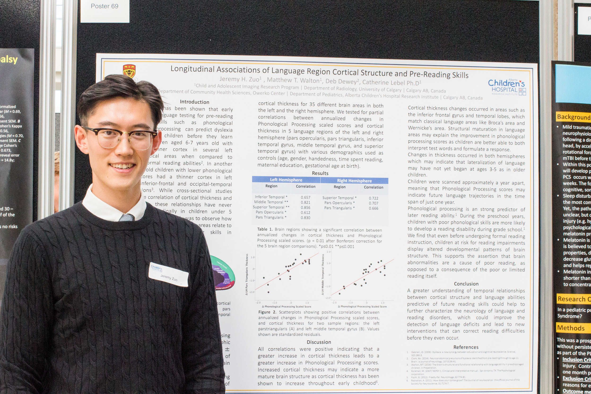
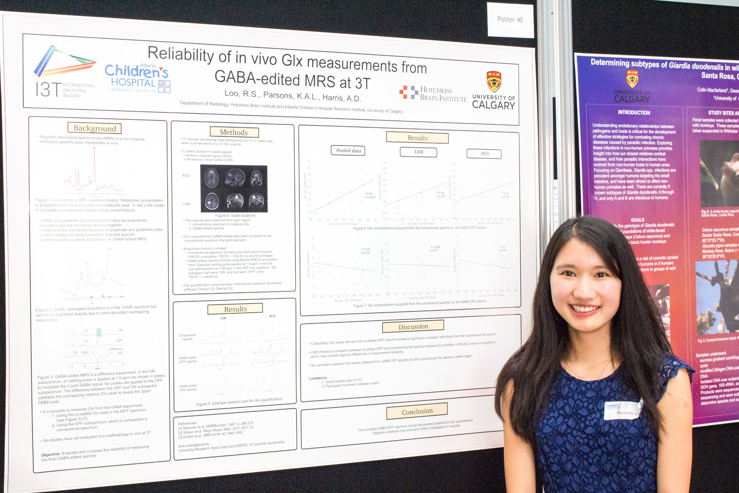
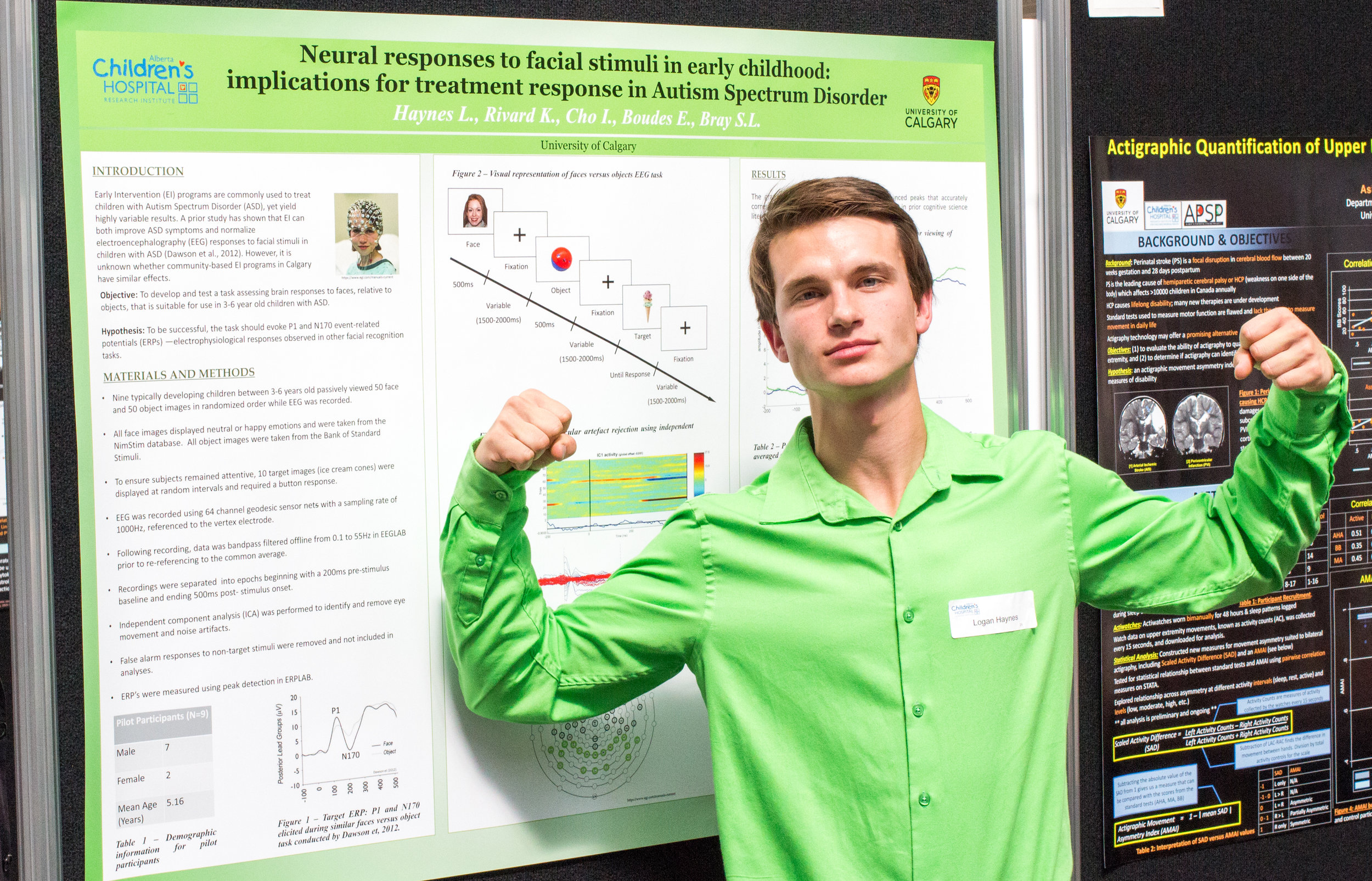
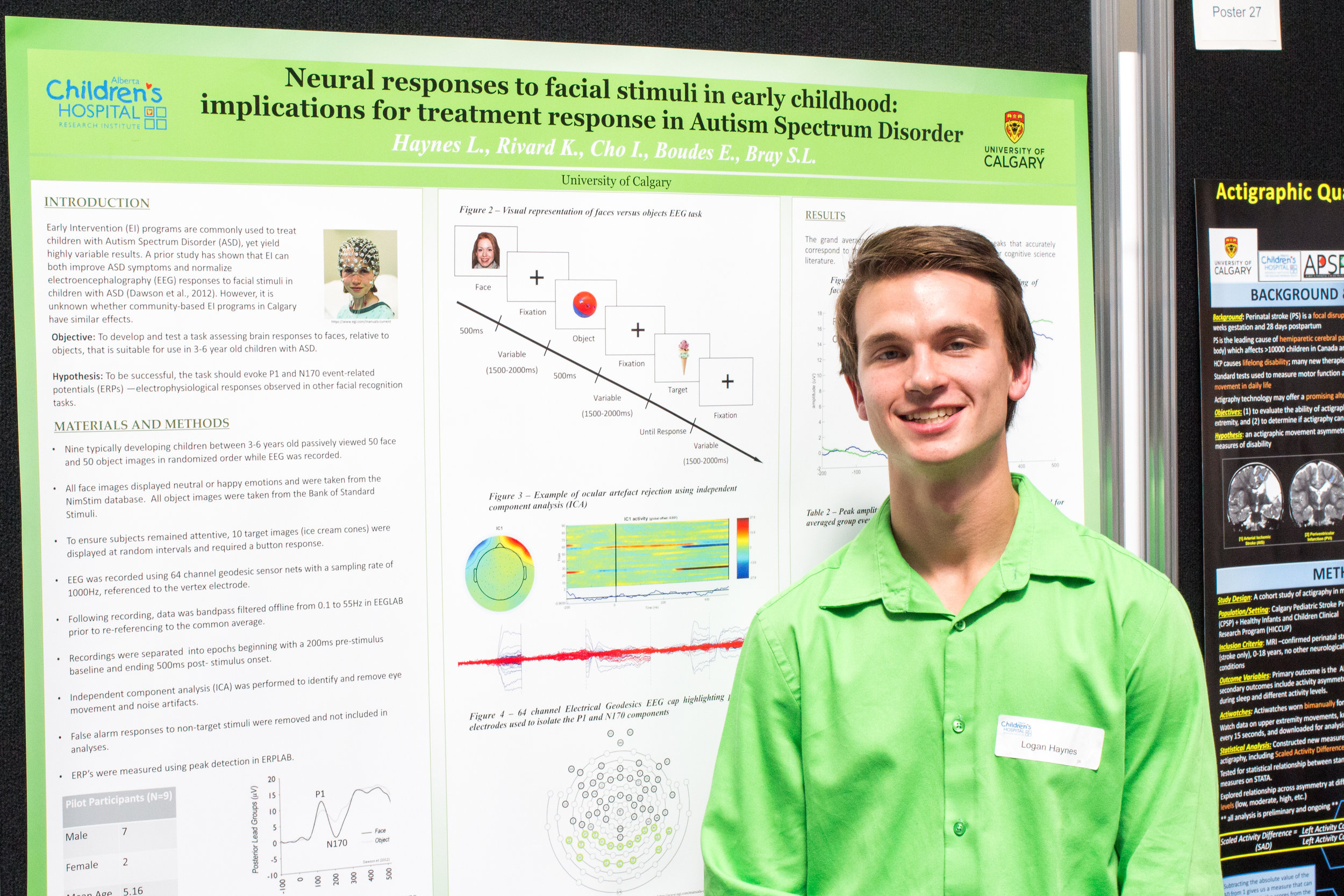
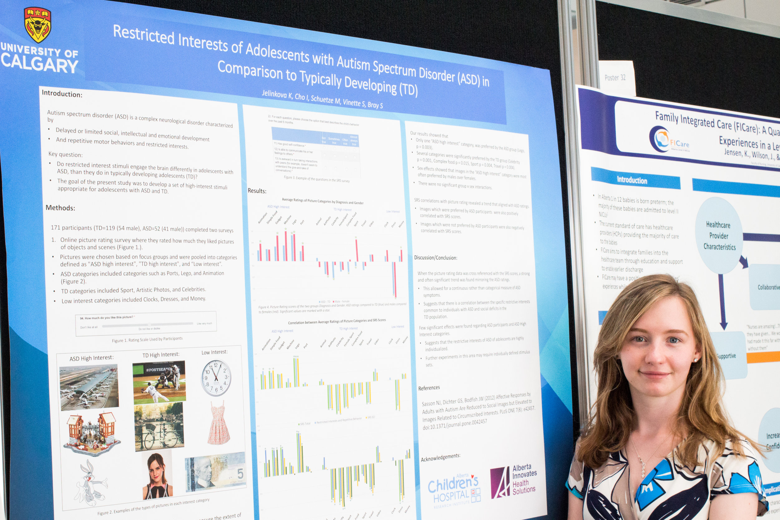
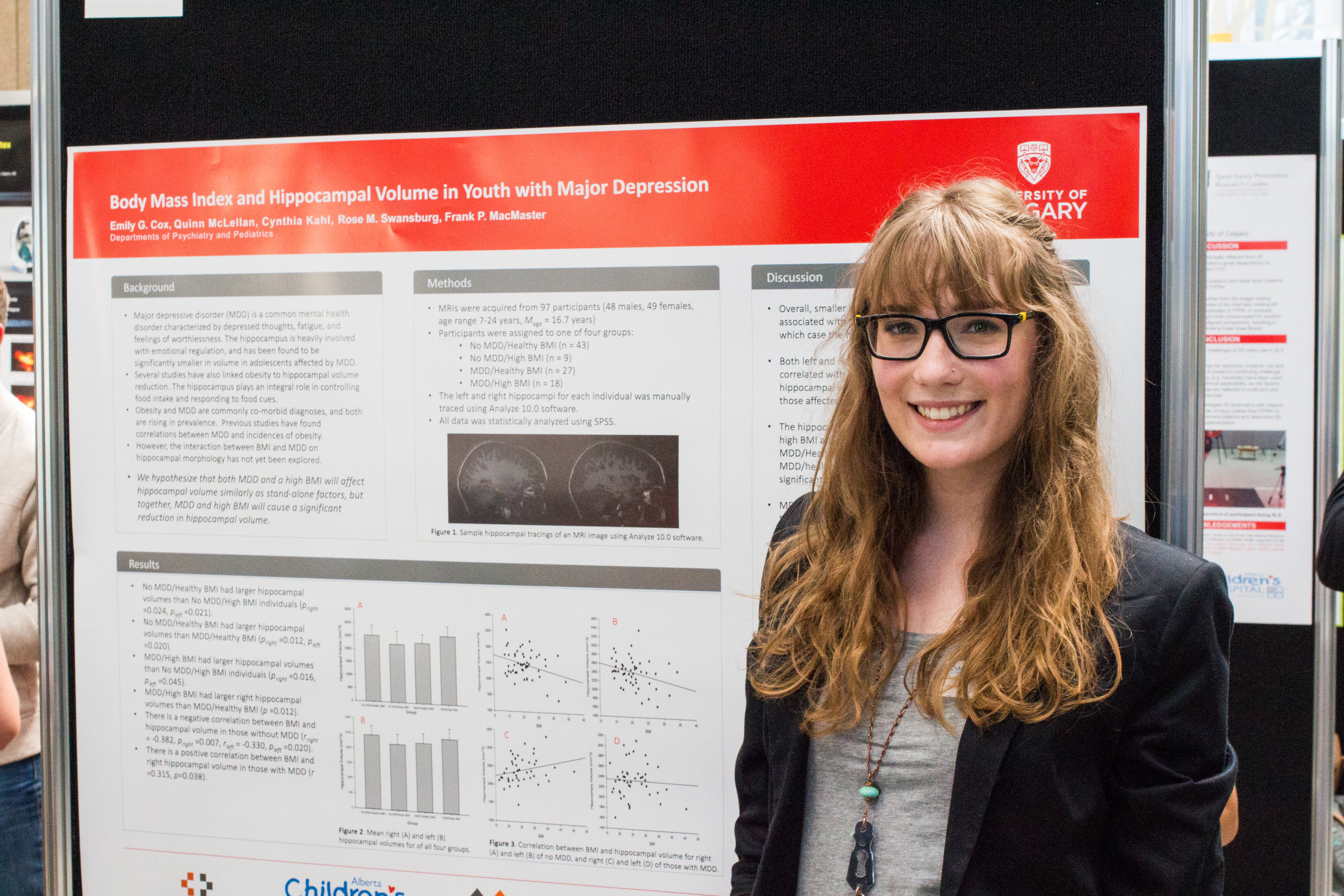
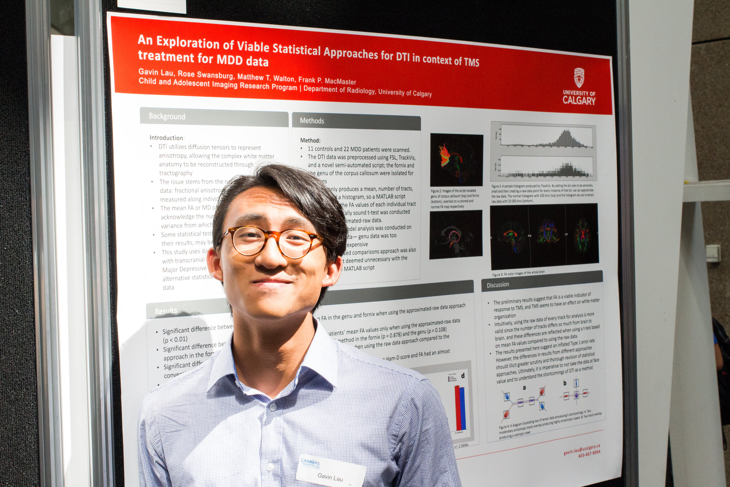
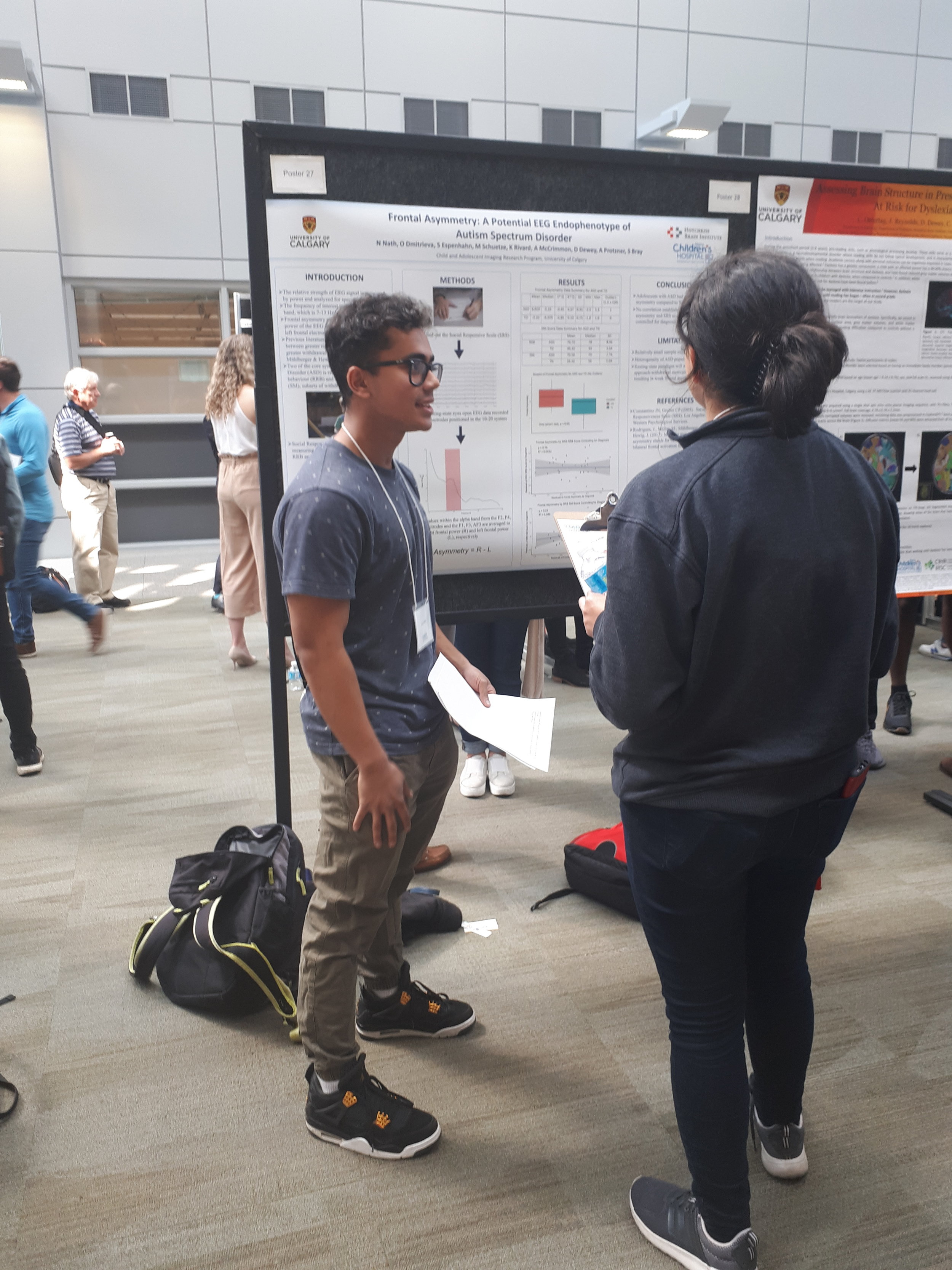
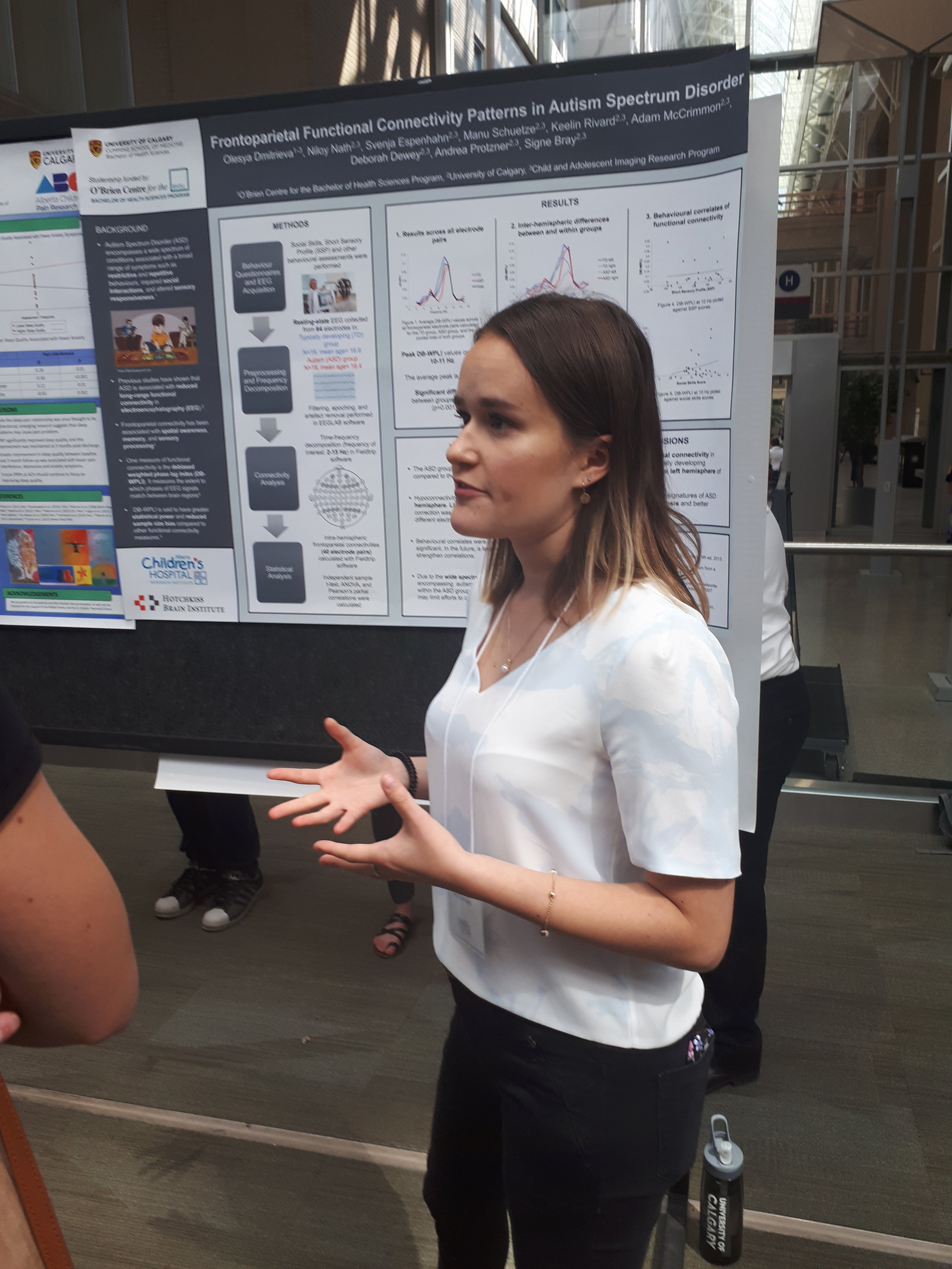
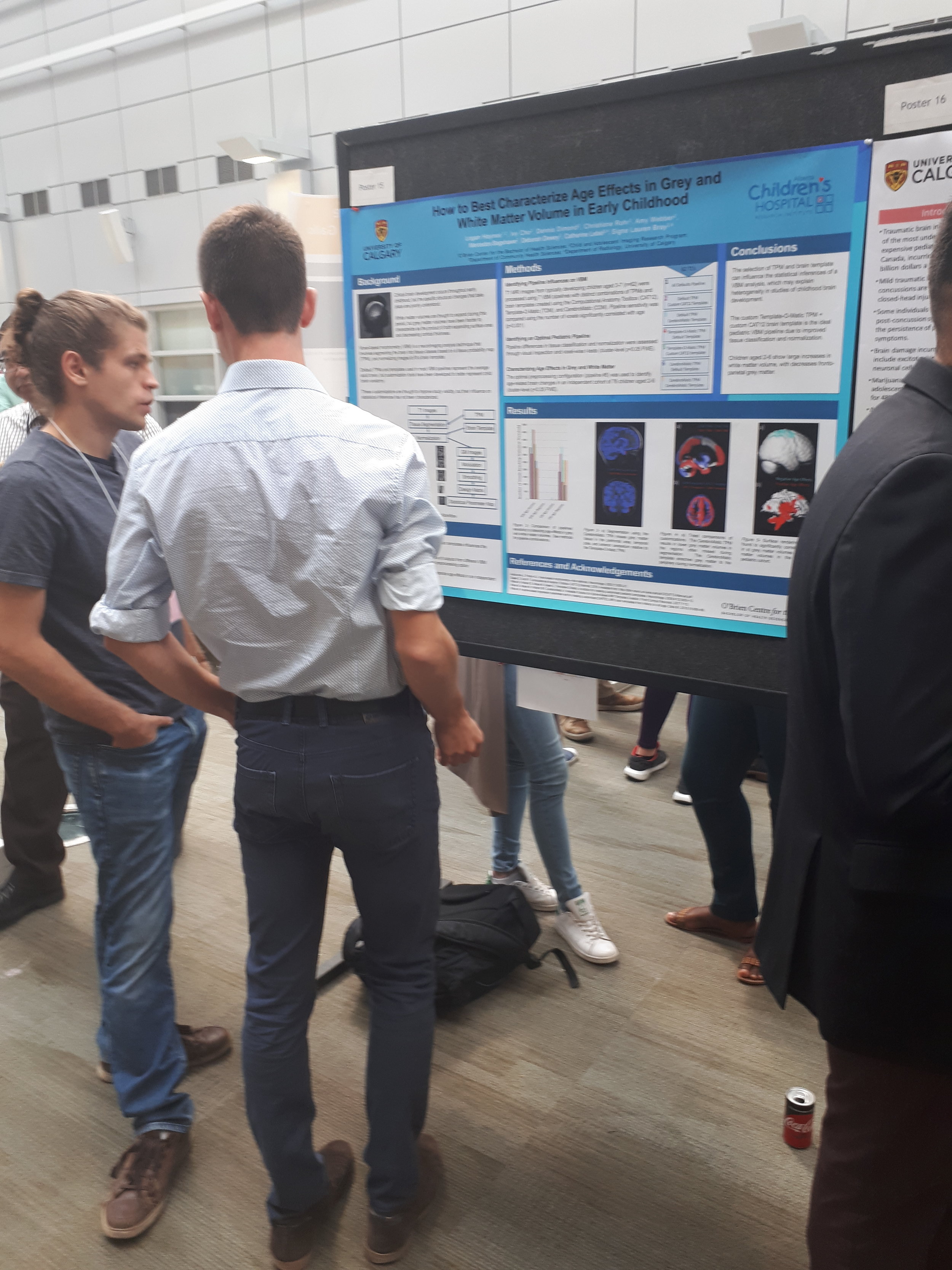
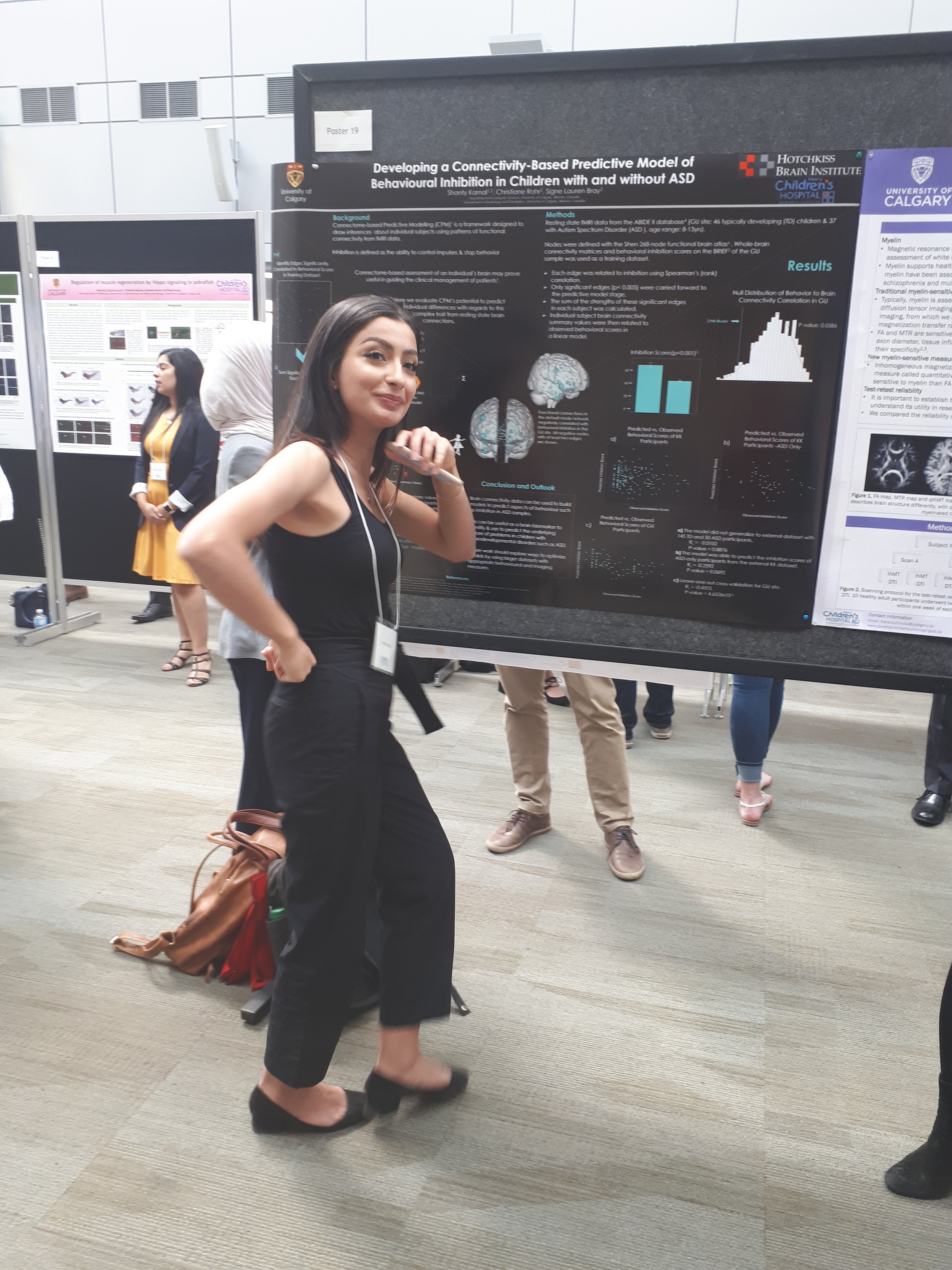
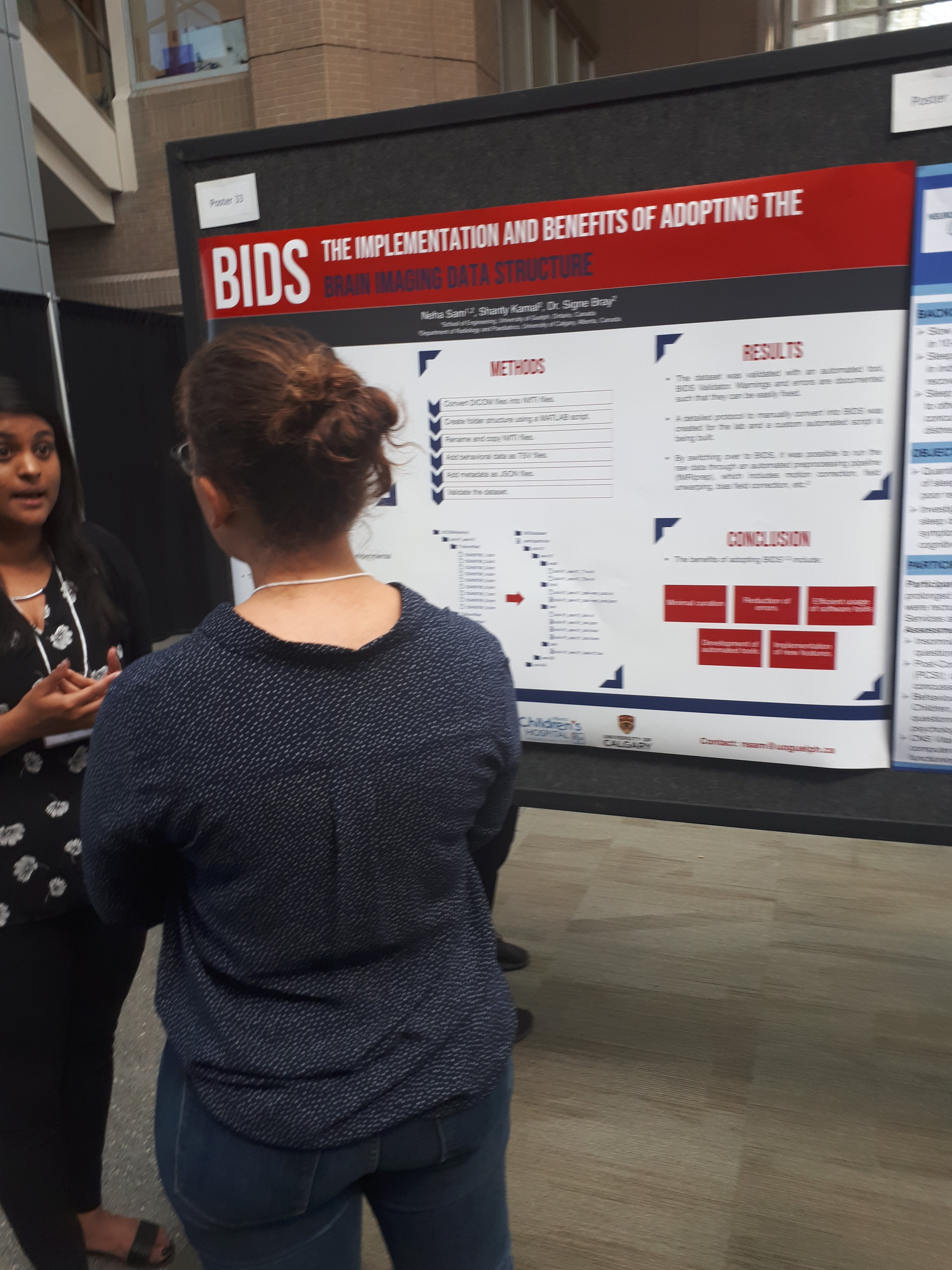
On some great work. Niloy Nath, Shanty Kamal, Olesya Dmitrieva, Neha Sam and Logan Haynes presented at the ACHRI and HBI summer student research days this week. Congratulations to Logan on runner up for best poster presentation!!

Posted
AuthorSigne Bray

Manu will be presenting part of her PhD project showing "Intact reinforcement learning with restricted interests in Autism Spectrum Disorder". Check out her poster on Tuesday, July 10, 09:30 - 13:00 at Poster Board F011.

Posted
AuthorSigne Bray

Both Manu and Michael from the lab were teamed up with a local artist who translated their work into a custom piece, presented at 'Your Brain on Art' -- http://www.branchoutfoundation.com/your_brain_on_art


While creating this piece I was thinking about what Manu and I had discussed in terms of how people on the autistic spectrum respond to the world: how they react positively to a variety of unanticipated stimulation, how they can become engrossed in a particular subject as well as the unique physical characteristics of each person's brain. From this I recognized similarities to artistic motivation; how certain visual sensations can be completely captivating: sunlight falling on a leaf, the shadow of fence across a sidewalk, the combination of colours on a canvas... From this, I allowed my own 'brain' to go wherever it pleased and this is what happened!


Kirsten Ringstrom, Artist (http://kirstenringstrom.org/)

Manu Schuetze, Neuroscientist

8x10 image in 13x17 frame

mixed media on raw canvas

John F. Gerrard

18 Unknowns

Print

Finished Size: 17" x 23"


Research Project: Gaze Behaviour & Emotional Perception in Schizophrenia - Michael Spilka, University of Calgary


When people hear schizophrenia, it’s often the positive symptoms such as delusions that come to mind. This study is focused on the recognition of emotion in faces, which is one of the social abilities affected in schizophrenia.  Michael Spilka and his team are studying the areas of the brain that are responsible for these social skills. Using fMRI and eye-tracking technology, they are hoping to find how the functioning of these brain regions are affected by how people look at faces, so they can further develop ways to help people with these issues.


The social ability to recognize which emotions are being conveyed is an important part of interacting with people. Schizophrenia can be an isolating illness, and I’m happy this part of the disease is being looked at.


I wanted to simulate what it might be like to take in a face and misrepresent it. I worked with images of “happy” people with the goal to make them ambiguous and eerie. I think it’s fascinating how a healthy brain can take visual data and interpret that into an abstract quality such as “happy”, or how there can be an unknown presented to the subject when things aren’t working properly. I learned from Michael that there isn’t one place in the brain that does this, rather different parts that work together. I also learned about how this process may be influenced by which parts of the face are focused on by the viewer. I find it fascinating how the seemingly mechanical nature of the brain can give rise to a unified subjective experience, and am intrigued by the mystery of how this works. My goal with this piece was to fail at interpreting the emotion. To strip it of any obvious signifier, leaving it abstract and undefined.

Posted
AuthorSigne Bray

Some of our lab members presented their research at the CAIR 5th Anniversary Research Symposium at the Alberta Children's Hospital on March 27, 2018. Congrats to everyone on their fantastic posters, and to Aneesh Khetani for winning Best Poster!

Aneesh Khetani - Evaluation of a visospatial n-back task in fMRI to examine working memory network activation in children following a mild traumatic brain injury - Winner of Best Poster!

Manu Schuetze - Intact reward functioning towards restricted interests in Austism Spectrum Disorder during reinforcement learning

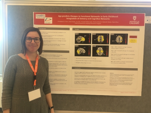
Christiane Rohr - Age predicts changes in functional networks in early childhood: Integration of sensory and cognitive networks

Logan Haynes - Exploring the effects of age-appropriate brain templates on voxel-based morphometry analysis

Posted
AuthorSigne Bray

Circumscribed interests in adolescents with Autism Spectrum Disorder can play an important role in forming new peer relationships based on shared interests. However, little is currently known about the circumscribed interests in adolescents with Autism Spectrum Disorder, and how it compares with those of typically developing adolescents. In our recently published article, we investigated the circumscribed interests of adolescents with Autism Spectrum Disorder.

Posted
AuthorSigne Bray

Early interventions for children with Autism are supported by the best evidence available to date and yet they don't guarantee to help every child that receives a diagnosis. Why do some children benefit from these interventions and others don't? This is an important question as not only are interventions expensive, but they also require a lot of time and effort from children and parents. 

In our new article, we reviewed the evidence for differences in learning and explore how learning strategies could be tailored to the unique abilities of children on the spectrum. We suggest that differences in treatment response may be due to differences in how individuals with ASD respond to, and learn form, reinforcers in the environment.


Posted
AuthorSigne Bray

We're excited to spend a few days in New Orleans at the Meeting of the International Neuropsychological Society. Ivy will be presenting her work on Frontostriatal Structural Connectivity in Autism Spectrum Disorder tomorrow, Thursday, February 2, 9:30-10:45 am (Poster # 58). 

Posted
AuthorSigne Bray

Dr. Signe Bray's work on brain development in children and adolescents is now online in Human Brain Mapping! Click here to read the abstract. 

Data sharing in neuroimaging allows groups from all over the world to make use of unique data sets. We have a new paper out today that used anatomical, arterial spin labeling and functional data from the 'Pediatric Template of Brain Perfusion' study (http://palgrave.nature.com/articles/sdata20153). We found that although there were similar declining age effects on gray matter volume and perfusion, the spatial patterns of these changes were largely independent. This means that multimodal imaging is necessary to get a clear and accurate picture of how the brain matures, and also that models linking changes in volume with changes in blood flow and function are more complex than one might expect.   

Posted
AuthorSigne Bray

Congratulations to Manu on winning 3rd place for best presentation at Neuron Night! She gave an interactive talk about her new research project: "The Effect of Transcranial Direct Current Stimulation on Reward Learning in Autism Spectrum Disorder."

Thank you to the Branch Out Neurological Foundation (http://www.branchoutfoundation.com/) for organizing a great event!


Posted
AuthorSigne Bray

Manu will be giving a talk about her current research on: "Neural correlates of variable learning in adolescents with ASD." You can hear her talk on November 15th from 1:00-2:45PM in SDCC 2 (Session #: 578). 

Keelin will be presenting a poster on: "Typical emotional processing of special interest stimuli in adolescents with Autism Spectrum Disorder." You can meet her on November 13th from 2:00-3:00PM. 

Posted
AuthorSigne Bray

...for winning this year's NSERC CREATE i3T Studentship and Queen Elizabeth II Graduate Master’s Scholarship! 

Posted
AuthorSigne Bray